
Development process of the prosthetic systems pipeline for chairside oral devices-two
2022-06-11
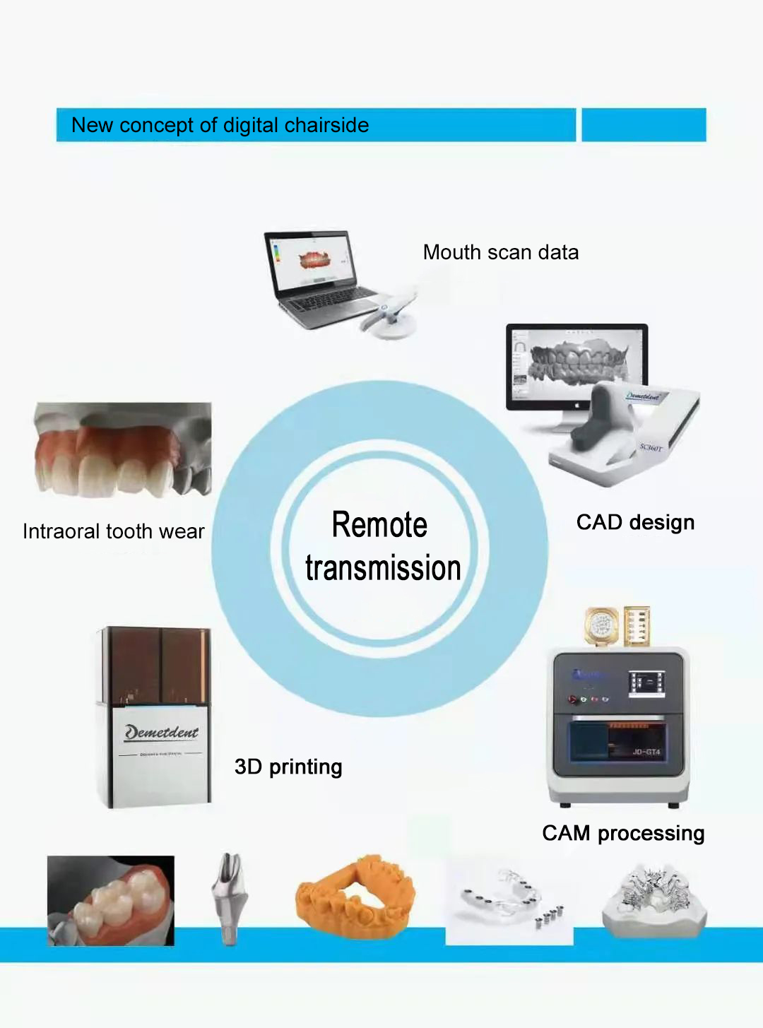
Chairside restorative systems mainly include digital intraoral scanning equipment, digital restorative design software (CAD), small CNC cutting equipment (CAM) and rapid sintering equipment.
The CEREC AC system, for example, consists of the following four main devices.
CEREC AC Intraoral Scanner: saves patients from the discomfort of traditional impression trays and uses the CEREC Omnicam, a small powder-free color camera, to acquire digital images; accurate natural color 3D images make scanning easier, more intuitive and more ergonomic.
CEREC CAD system: generates excellent repair recommendations based on the unique "Biojaw" function, saving time and providing the best repair design as soon as possible; benefits from a simple, well-structured and intuitive user interface.
CEREC CAM system: optimally synchronized with the CEREC software, very precise, with smooth surfaces and edges and very fine restorations, regardless of the material chosen.
CEREC Rapid Sintering Equipment: Fully contoured zirconia restorations are sintered and glazed in the compact CEREC Rapid Sintering Furnace. The induction technology allows for the shortest possible sintering and wear times and is very intuitive and simple to use. In addition to zirconia, CEREC Rapid Sintering can process all other types of glass crystals.
There are two types of techniques for obtaining digital impressions of the oral cavity: indirect and direct methods. The indirect method is the more maturely applied 3D scanning technology for dental plaster models, and the dental model scanner has been more widely used clinically as a regular configuration in modern digital technician centers. The direct method is the fast-developing intraoral digital impression technology (Interral Scanner), which applies a small probing optical scanning probe to obtain the surface morphology of teeth, gums and other soft and hard tissues directly in the patient's mouth, omitting the operation of making impressions and turning plaster models, bringing a rather comfortable consultation experience for patients.
The existing intraoral scanning techniques can be classified according to the technical principles as.
At present, more than ten sets of intraoral scanning systems have been released internationally, including 3Shape (Denmark), iTero (USA), Lythos (USA), Cerec (Germany), 3M Lava (USA), Dental Wings (iJI take big), Planmeca (Finland), etc. The scanning efficiency of each system does not vary much (single tooth position The scanning efficiency of each system is not very different (15-20S for a single tooth position), and most of them have realized data opening under free or paid conditions. At present, Guangdong CADCAM has also launched its intraoral scanning system in China.
The intraoral scanner can realize the direct transformation from intraoral dentition to digital dental model, avoiding the errors in the process of impression making, transmission and plaster model instillation, and the color 3D scanning technology can restore the real 3D morphology and texture color of oral tissues to the maximum extent, with obvious advantages of flexibility and intuitiveness, and good prospects for clinical application.
The CEREC AC system, for example, consists of the following four main devices.
CEREC AC Intraoral Scanner: saves patients from the discomfort of traditional impression trays and uses the CEREC Omnicam, a small powder-free color camera, to acquire digital images; accurate natural color 3D images make scanning easier, more intuitive and more ergonomic.
CEREC CAD system: generates excellent repair recommendations based on the unique "Biojaw" function, saving time and providing the best repair design as soon as possible; benefits from a simple, well-structured and intuitive user interface.
CEREC CAM system: optimally synchronized with the CEREC software, very precise, with smooth surfaces and edges and very fine restorations, regardless of the material chosen.
CEREC Rapid Sintering Equipment: Fully contoured zirconia restorations are sintered and glazed in the compact CEREC Rapid Sintering Furnace. The induction technology allows for the shortest possible sintering and wear times and is very intuitive and simple to use. In addition to zirconia, CEREC Rapid Sintering can process all other types of glass crystals.
Digital intraoral scanning devices
There are two types of techniques for obtaining digital impressions of the oral cavity: indirect and direct methods. The indirect method is the more maturely applied 3D scanning technology for dental plaster models, and the dental model scanner has been more widely used clinically as a regular configuration in modern digital technician centers. The direct method is the fast-developing intraoral digital impression technology (Interral Scanner), which applies a small probing optical scanning probe to obtain the surface morphology of teeth, gums and other soft and hard tissues directly in the patient's mouth, omitting the operation of making impressions and turning plaster models, bringing a rather comfortable consultation experience for patients.
The existing intraoral scanning techniques can be classified according to the technical principles as.
①Triangulation imaging techniques (stereovision and triangulation).
The basic principle is that the beam emitted from the light source is projected onto the tooth surface and imaged on the charge coupled device (CCD) after reflection. The three-dimensional coordinate information (XYZ) of the measured point on the tooth can be obtained by solving the similar triangle of the optical system through the known object distance, image distance, angle between the main optical axis and the CCD imaging plane, angle between the incident light and the main optical axis, and the imaging position information of the corresponding image point on the CCD.②Confocal microscopy.
The point light source and point detection are achieved by using an illumination pinhole placed behind the light source and a detection pinhole placed in front of the detector. The light emitted from the illumination pinhole is focused on a point in the focal plane of the tooth, and the reflected light from this point is returned by the original light path and imaged in the probe pinhole, while the reflected light in the non-focused range is blocked by the probe pinhole and not imaged, thus obtaining only the morphological data of the tooth in the focal plane. The illumination pinhole and the probe pinhole are conjugate to the irradiated or probed point, so the probed point is the confocal point and the plane where the probed point is located is the confocal plane (focal plane for short). By scanning layer by layer, the focal plane morphology data at different depths of the tooth are obtained, and thus the three-dimensional morphology of the tooth is constructed.③Active wavefront sampling.
By using the rotating eccentric aperture device set in the sampling optical path, the reflected light from the measured point on the tooth is filtered and a defocused image with circular trajectory is formed in the imaging plane, and the spatial coordinates of the measured point on the tooth surface are calculated by measuring the radius of the defocused image and combining with the known parameters of the optical path system. The light filtering effect of the eccentric aperture device can well prevent the images of different areas of the tooth surface from overlapping, thus improving the spatial resolution of the images.At present, more than ten sets of intraoral scanning systems have been released internationally, including 3Shape (Denmark), iTero (USA), Lythos (USA), Cerec (Germany), 3M Lava (USA), Dental Wings (iJI take big), Planmeca (Finland), etc. The scanning efficiency of each system does not vary much (single tooth position The scanning efficiency of each system is not very different (15-20S for a single tooth position), and most of them have realized data opening under free or paid conditions. At present, Guangdong CADCAM has also launched its intraoral scanning system in China.
The intraoral scanner can realize the direct transformation from intraoral dentition to digital dental model, avoiding the errors in the process of impression making, transmission and plaster model instillation, and the color 3D scanning technology can restore the real 3D morphology and texture color of oral tissues to the maximum extent, with obvious advantages of flexibility and intuitiveness, and good prospects for clinical application.
 CNC milling machine operation need to pay attention to what?
 - What is orography?
- What is a denture?
- What is a CNC milling machine?
 Application and development of chairside digital systems in implant restorations
  Oral chairside device prosthetic system industry development process3
  Oral chairside device restorative system industry development process one
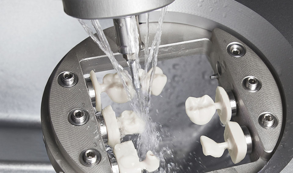
  CNC milling machine on the teeth: grinding machine
  ## what's a dental milling machine?
  Your satisfaction is our ultimate goal